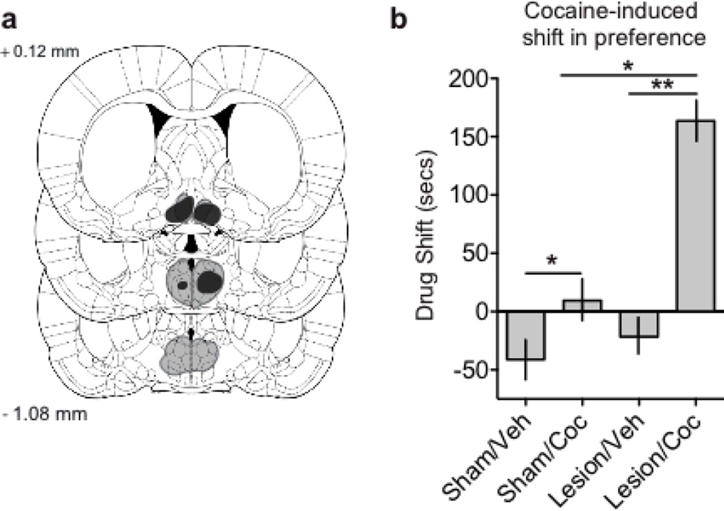
Figure 3.
Lesions of the mPOA increased cocaine-induced conditioned place preference. (a) Representative perimeters of smallest (black) and largest (gray lines) lesion for animals with radiofrequency lesions of their mPOA, drawn from Paxinos and Watson (2007). (b) Graph demonstrating the shift in chamber preference across treatment conditions, cocaine treated animals displayed a cocaine-induced shift in time spent in an initially non-preferred chamber, whereas animals receiving lesions of the mPOA displayed an even greater shift. Values are expressed as mean ± SEM; * p < 0.05, ** p < 0.01. Coronal plates were adapted from The Rat Brain in Stereotaxic Coordinates (6th ed.), Fig 3a from pages 75, 80, and 84, by G. Paxinos & C. Watson, 2007, New York, NY: Academic Press. Copyright 2007 by Elsevier Academic Press; adapted with permission.

