Figure 1.
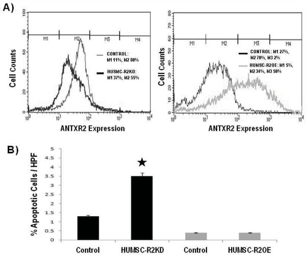
ANTXR2 is expressed on human uterine smooth muscle cells (HUSMC) and knockdown of ANTXR2 increased rates of HUSMC apoptosis. A) These graphs represent results from one of two flow cytometry assessments that were performed. Flow cytometry confirmed the presence of ANTXR2 protein on the surface of HUSMC. Marker 1 (M1) represents the ANTXR2 negative cells and Marker 2 and 3 (M2, M3) represents the CMG2 positive cells. On average, we were able to achieve approximately 40% lentiviral-mediated knock down (KD) and 30% retroviral-mediated over-expression (OE) relative to control (CTL). B) TUNEL (TdT mediated dUTP Nick End Labeling) staining results showed lentiviral-mediated KD of ANTXR2 in HUSMC increased apoptosis whereas retroviral-mediated OE of ANTXR2 did not affect the number of apoptotic cells.

