
Figure 3.
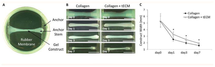
Contraction of constructs. (A) Top view of Tissue Train culture plate together with gel construct shows that the cell-collagen construct was fixed at each end by bonding to the nylon anchor stem. (B) Photos were taken to record the change of construct width. Reduced contraction is seen in the tECM-supplemented collagen constructs (right panels) compared to the collagen gel contructs (left panels). (C) The average width of the tECM-supplemented constructs is significantly higher than that of pure collagen constructs at days 1, 3 and 7. Scale bar=10 mm; * indicates p<0.05, n=6.