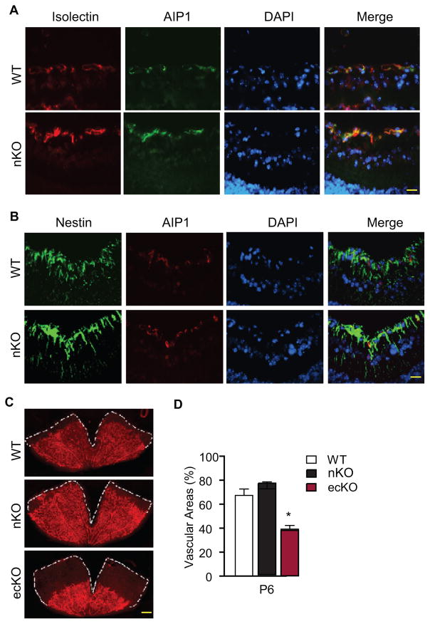
Figure 3. Effect of a neuronal-specific AIP1 deletion in retinal vascular development.
P6 retinas from WT (AIP1lox/lox), the AIP1-nKO (AIP1lox/lox:Nestin-Cre) and the AIP1-ecKO (AIP1lox/lox:Tie2-Cre) were collected. A–B. AIP1 expression was detected by immunostaining of cross sections by anti-AIP1 together with isolectin staining (for EC) or nestin staining (for neuronal cells).). Scale bar: 50 μm. C–D. The superficial retinal vasculatures were visualized by isolectin staining (C). Scale bar: 500 μm. Vascularized areas were quantified as a percentage of the total retinal surface (D). n=10 retinas from 5 mice for each strain. *, p<0.05 comparing AIP1-ecKO to WT. No significance was detected comparing AIP1-nKO to WT group.

