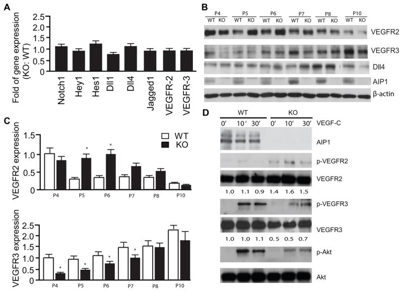
Figure 4. VEGFR-3, but not VEGFR-2 or Notch signaling, is specifically reduced in AIP1-KO retinas.
A. Retinas from WT and AIP1-KO at P5 were collected. Gene expression of VEGFR-2, VEGFR-3 and Notch signaling molecules were determined by qRT-PCR. Data represent fold changes by taking WT as 1.0. Data are mean ± SEM from 6 retinas for each group. B–D. Protein expression of VEGFR-2, VEGFR-3 and Notch signaling molecules. Retinas from WT and AIP1-KO at P4-P10 were harvested. Protein expressions were determined by immunoblotting with respective antibodies. Representative blots from four independent experiments are shown in B. Relative levels of VEGFR-2 and VEGFR-3 expression are represent by taking WT P4 as 1.0. Data are mean ± SEM from 6 retinas for each group. *, p<0.05 comparing AIP1-KO to WT. D. Effects of AIP1 deletion on VEGF-C signaling. Retinal EC were isolated from P7 retinas and were serum-starved overnight. Cells were treated with VEGF-C (100 ng/ml) for indicated times. Phosphorylations of VEGFR-2, VEGFR-3 and Akt were determined by Western blot with phosphor-specific antibodies. Total protein levels of VEGFR-2, VEGFR-3, Akt and AIP1 were determined by Western blot with respective antibodies. Relative levels of VEGFR-3 and VEGFR-2 are indicated below the blot with untreated WT group as 1.0. Similar results were obtained from additional two experiments.

