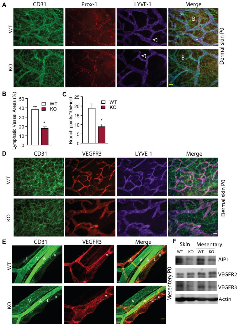
Figure 5. AIP1 deletion delays lymphangiogenesis in neonatal skin and mesentery.
Dermal skin and mesentery from WT and AIP1-KO at P0 were collected. A. Blood and lymphatic vessels in dermal skin were visualized by whole-mount immunostaining with anti-CD31 (green), anti-Prox-1 (red) and anti-LYVE-1 (purple). Merged images are shown on the right. Blood and lymphatic vessels as well as lymphatic vessel branches are indicated. Scale bar: 50 μm. B–C. % of lymphatic vessel areas (B) and lymphatic vessel branches (C) were quantified. n=3 sections from 3 mice for each strain. *, p<0.05 comparing AIP1-KO to WT. D. Blood and lymphatic vessels in dermal skin were visualized by whole mount immunostaining with anti-CD31 (green), anti-VEGFR-3 (red) and anti-LYVE-1 (purple). Merged images are shown on the right. Scale bar: 50 μm. E. Representative whole-mount immunostaining of mesenteric vessels with anti-CD31 (green), anti-Prox-1 (red) and anti-LYVE-1 (purple). Merged images are shown on the right. A: artery; V: vein; L: lymphatics. The lymphatic valves are indicated by *. Scale bar: 50 μm. F. Protein expression of VEGFR-3. Skin and mesentary tissues from WT and AIP1-KO at P0 were harvested. Protein expressions were determined by immunoblotting with respective antibodies. Representative blots from four independent experiments are shown.

