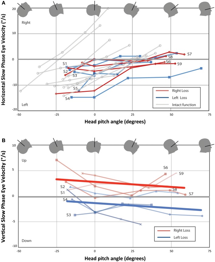
Figure 2.
Change in nystagmus slow-phase horizontal (A) and vertical (B) eye velocity from baseline eye velocity for all UVH subjects. Each point represents the difference in average SPV between a 40-s time interval once the subject has entered the bore and baseline average SPV in darkness prior to entry. Right-sided UVH subjects are labeled to the right and left-sided UVH subjects to the left of each line. Ten subjects with bilateral intact vestibular function from Roberts et al. are plotted in (A) for comparison (3). Lines of best-fit (linear least-squares method) are shown in (B) for right-sided UVH and left-sided UVH. (A) All subjects developed a slow-phase left nystagmus that decreased in velocity with chin down head pitch, with four subjects demonstrating reversal of horizontal nystagmus direction with head pitch beyond a null position. (B) Right-sided loss subjects developed slow-phase-up nystagmus inside the magnetic field and left-sided loss subjects developed slow-phase-down nystagmus.

