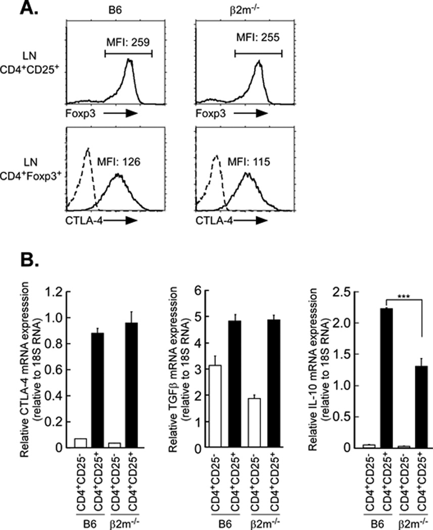
Figure 8. MHC class I expression enhances IL-10 but not CTLA-4 and TGF-β.

A. MHC class I deficiency does not affect Foxp3 and CTLA-4 expression in Tregs. Lymph node T cells were first stained for surface CD25, fixed and then stained for Foxp3 and CTLA-4. Top panels display Foxp3 intracellular staining of gated CD4+CD25+ LNT cells and the numbers indicate the MFI of Foxp3. Bottom panels display intracellular CTLA-4 staining of gated CD4+Foxp3+ LNT cells and the numbers indicate the MFI of CTLA-4. Dashed lines represent negative control staining. Data are representative of three independent experiments.
IL-10 RNA is reduced in β2m−/− CD4+CD25+ T cells relative to that in B6 CD4+CD25+ T cells. Total RNA was isolated from purified CD4+CD25− Tconv and CD4+CD25+ Tregs and subjected to quantitative RT-PCR for CTLA-4, TGF-β and IL-10. 18S rRNA was used as an internal control. Mean ± SEM of triplicate samples in three experiments. ***p<0.001.
