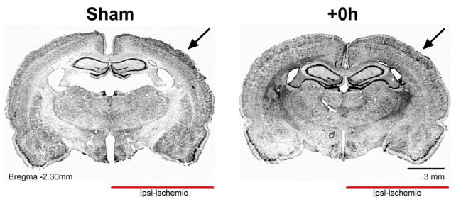
Figure 5.

Cortical structure in +0h subjects remains equivalent to surgical shams at 4 months post-pMCAO. Representative coronal sections showing primary somatosensory cortex in sham and +0h subjects. Arrows point toward MCA blood supply territory for this cortical region. Cresyl violet staining shows no glial scarring in +0h subjects.
