
Figure 1.
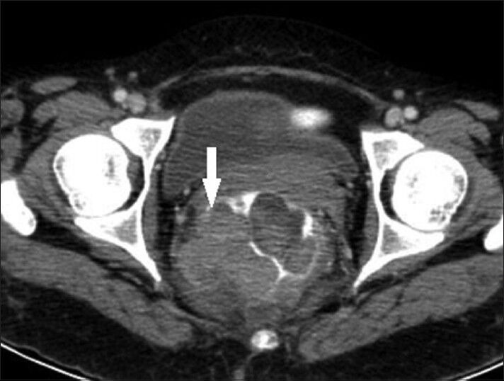
43-year-old woman with protruding anal lesion diagnosed with melanoma of the rectum. Contrast-enhanced axial CT scan images of pelvis shows heterogeneous mass lesion in rectum with extension into mesorectal fat (white solid arrow).
Official websites use .gov
A
.gov website belongs to an official
government organization in the United States.
Secure .gov websites use HTTPS
A lock (
) or https:// means you've safely
connected to the .gov website. Share sensitive
information only on official, secure websites.
43-year-old woman with protruding anal lesion diagnosed with melanoma of the rectum. Contrast-enhanced axial CT scan images of pelvis shows heterogeneous mass lesion in rectum with extension into mesorectal fat (white solid arrow).