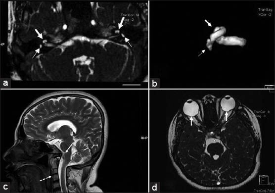
Figure 3.

27-year-old female with poor vision and inability to hear was subsequently diagnosed with CHARGE syndrome. (a) Axial Constructive Interference in Steady State (CISS) T2-weighted MRI at the level of internal auditory meatus shows bilateral hypoplasia of semicircular canals seen as small saccular structures instead of normal “D” like canalicular structure (small arrows) and cochlea shows only one and half (11/2) turns instead of normal two and half (21/2) or more (large arrows). (b) Three-dimensional reconstruction of CISS MRI images of the right internal auditory apparatus confirming rudimentary semicircular canals (small arrow) and hypoplasia of cochlea (large arrow). (c) T2 Fast spin echo mid-sagittal reconstruction of MRI head shows associated craniovertebral junction anomaly with fused C2 and C3 vertebrae with rudimentary intervertebral disc (dotted arrow) and basilar invagination (solid arrow). (d) Axial CISS T2 weighted MRI at the level of orbits shows defects of choroid (solid arrows).
