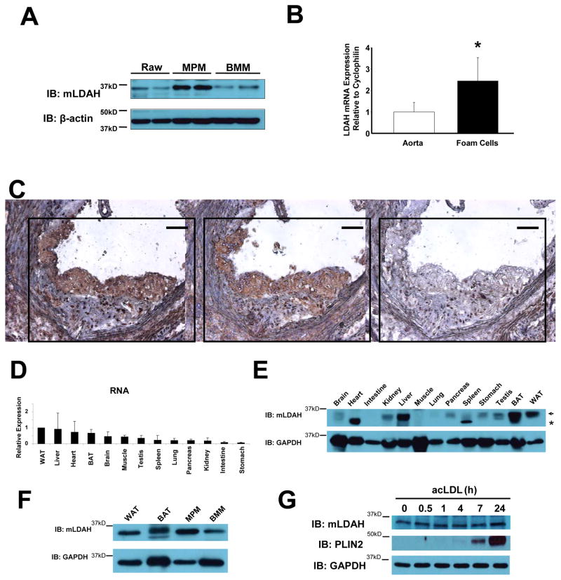
Figure 5. LDAH expression in mouse tissues.
(A) Immunoblot with anti-mLDAH in lysates of RAW 264.7 macrophages, thioglycollate elicited MPM, and mouse BMM. (B) qPCR analysis comparing LDAH levels in RNA isolated by LCM from macrophage/foam cell rich-areas within atherosclerotic lesions and whole aorta RNA of apoE−/− mice. Data are shown as mean ± SD. (n=5). *p<0.05. (C) Immunoperoxidase staining with mLDAH antisera (left panel, brown color) and anti-Lamp-2 (macrophage marker, middle panel, brown color) in consecutive sections of the aortic sinus of apoE−/− mice. Preimmune serum was used as negative control (right panel). Bar= 100 μM. (D) qPCR analysis of LDAH RNA levels in male mouse tissues. (n=3). (E) Immunoblotting with anti-mLDAH in mouse tissues. The arrow indicates the specific mLDAH band. *non-specific band. (F) Immunoblotting with anti-mLDAH comparing mLDAH levels between WAT, BAT, MPM and BMM. (G) LDAH levels were not affected by cholesterol loading with acLDL (50 μg/ml in DMEM-1% FBS). The LD-associated protein PLIN2 was highly induced by the treatment.

