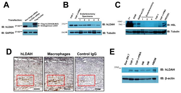
Figure 6. LDAH is expressed in foam cells within human atherosclerotic lesions.
(A) The specificity of the antibody generated against hLDAH was tested in HEK293 cells transfected with hLDAH siRNA and overexpressing flag-hLDAH. (B) 15 μg of THP-1 lysates and 30 μg of human endarterectomy lysates were used to assess hLDAH expression by immunoblotting. The figure displays five representative examples from a total of thirteen endarterectomy specimens analyzed; hLDAH was detected in all the samples tested. (C) Immunoblotting with anti-HSL on 70 μg of protein lysates from mouse RAW 264.7 macrophages, human THP-1 macrophages, HEK 293 cells, and endarterectomy specimens. HEK293 cells transfected with HSL were used as a positive control (C+). (D) Immunoperoxidase staining with anti-hLDAH (left panel, brown color) and anti-CD68 (macrophage marker, middle panel, brown color) in consecutive sections of human endarterectomy specimens. Rabbit IgG was used as negative control (right panel). Bar= 100 μM. (E) Immunoblotting with anti-hLDAH on 7.5 μg of protein lysates from RAW 264.7 macrophages, THP-1 macrophages, human monocytes (HM) and human monocyte-derived macrophages (HMDM).

