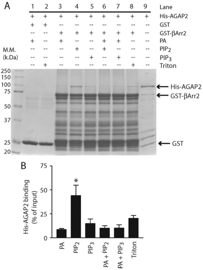
Figure 2. PtdIns(4,5)P2 enhanced the interaction between AGAP2 and β-arrestin2.

(A) Effect of phospholipids on AGAP2 binding to β-arrestin2 (βArr2). Purified His6–AGAP2 (150 nM) was incubated with GST–β-arrestin2 (500 nM) at room temperature for 1 h in the presence or absence of phospholipids at the following concentrations: PA, 360 μM; PtdIns(4,5)P2 (PIP2), 45 μM; PtdIns(3,4,5)P3 (PIP3), 10 μM; presented in micelles with 0.1% Trition X-100. Glutathione beads were washed three times and bound proteins were resolved by SDS/PAGE (10% gel) and visualized by Coomassie Blue staining. Molecular mass (M.M.) is shown on the left-hand side of the Western blot in kDa. (B) Quantification of His6–AGAP2 binding to GST–β-arrestin2. The binding assay was repeated three times and analysed by densitometry. *P < 0.05 compared with Triton X-100.
