
Figure 1.
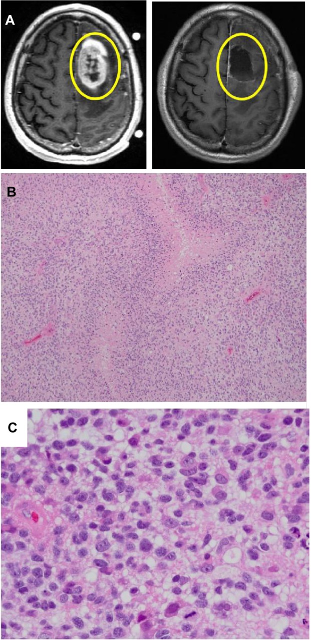
Radiographic (MRI) images and histology of GBMs.
Notes: (A) shows an MRI of a GBM before (left) and after (right) surgery. Yellow circles show the area of tumor (left) and the resection cavity following surgery (right). (B) (low power, 100×) and (C) (high power, 600×) are hematoxylin/eosin stains of a section of a GBM used in histopathologic diagnosis. (B) shows the typical hypercellularity, cytological atypia, and prominent pseudopalisading necrosis of a GBM. (C) at higher power (same tumor, different section), better illustrates the cellular atypia and mitotic activity in the GBM.
Abbreviations: GBM, glioblastoma multiforme; MRI, magnetic resonance imaging.