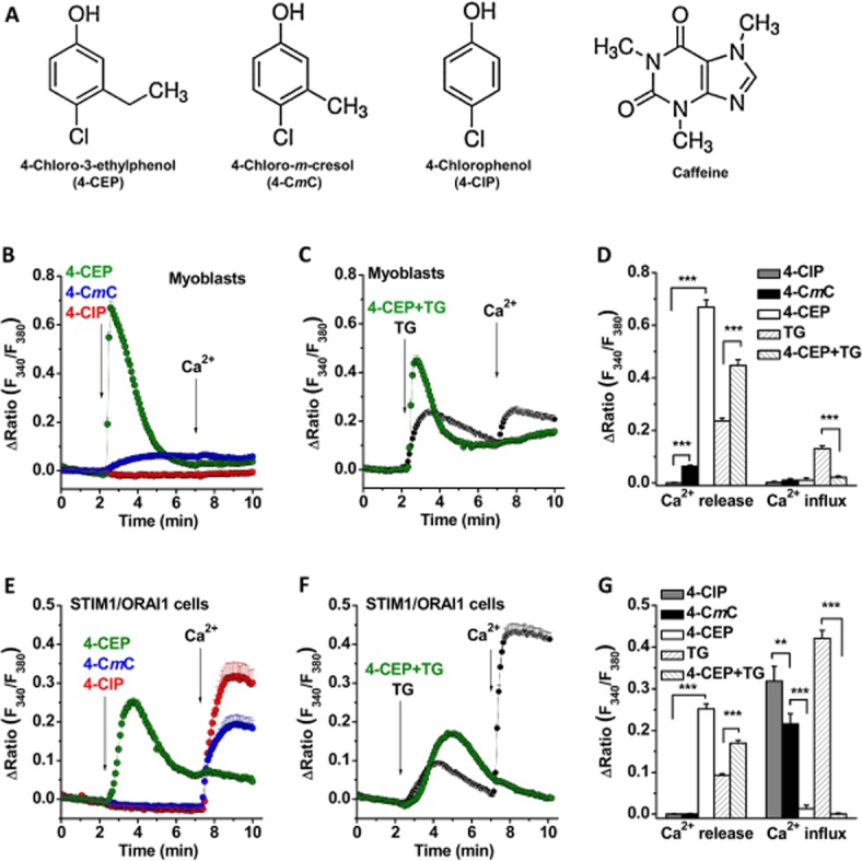
Figure 1.

4-CEP induced Ca2+ release but abolished store-operated Ca2+ entry in rat L6 myoblasts and HEK293 cells co-expressing STIM1/ORAI1. (A) Structures for 4-CEP, 4-CmC, 4-ClP and caffeine. (B) The effects of 4-CEP, 4-CmC and 4-ClP on the cytosolic Ca2+ level in rat L6 myoblasts. The cells were maintained in Ca2+ free solution first and then perfused with different compounds (500 μM for each compound) in Ca2+ free and 1.5 mM Ca2+ solution as indicated by the arrows. (C) 4-CEP (500 μM) nearly abolished the TG (1 μM)-induced Ca2+ influx in the myoblasts. (D) Mean ± SEM data for the comparison of Ca2+ release and influx induced by 4-ClP, 4-CmC, 4-CEP and TG in the myoblasts (n = 19–40 cells in each group). (E) The effects of 4-CEP, 4-CmC and 4-ClP (500 μM) in STIM1/ORAI1 cells. (F) 4-CEP (500 μM) abolished the TG (1 μM)-induced Ca2+ influx in STIM1/ORAI1 cells. (G) Mean ± SEM data for Ca2+ release and influx induced by 4-ClP, 4-CmC, 4-CEP and TG in the STIM1/ORAI1 cells (n = 18–26 cells in each group). Ca2+ release was measured as the peak of Ca2+ release signal; and Ca2+ influx was measured at 1 min after Ca2+ re-addition. One-way ANOVA with Bonferroni test was used for (D) and (G). ** P < 0.01, *** P < 0.001.
