
Figure 2.
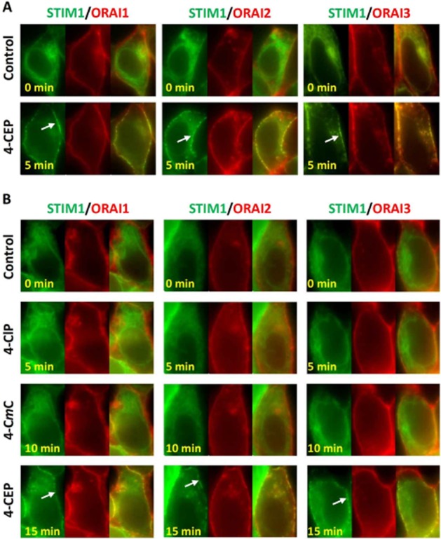
STIM1 translocation and clustering induced by 4-CEP, but not by 4-CmC and 4-ClP in the STIM1/ORAI1-3 cells. (A) Application of 500 μM 4-CEP for 5 min induced persistant STIM1-EYFP puncta (green, indicated by arrow) at the plasma membrane in STIM1-EYFP/mCFP-ORAI1-3 cells, while cytosolic clustering of STIM1 was not evident. The mCFP-ORAI fluorescence was converted into red pseudocolour in the pictures. The merged images are shown on the right of each group. The example cell for each group is a representative of 15 to 26 cells. (B) The cells were successively challenged with 500 μM 4-ClP, 500 μM 4-CmC and 500 μM 4-CEP in 1.5 mM Ca2+ solution with 5 min interval for each drug. STIM1 puncta at the plasma membrane (arrow) seen in 4-CEP-treated cells, but no STIM1 clustering was induced by 4-CmC and 4-CIP.