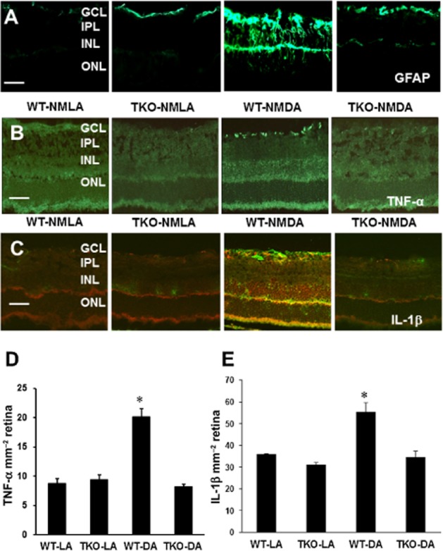
Figure 3.

TXNIP deletion inhibits Müller cell activation and retinal inflammation. (A) Representative images showing a substantial increase in GFAP immunoreactivity in the filaments of Müller cells spanning across the inner portion of the neural retina in WT-NMDA mice as compared with NMLA controls. (B, D) Representative images and statistical analysis of prominent immunolocalization of TNF-α in the GCL and INL in WT-NMDA mice as compared with NMLA controls. WT-NMDA mice had ∼2-fold increase in TNF-α expression relative to all other groups. (C, E) Representative images and statistical analysis of immunolocalization of IL-1β (red) and GFAP (green) in NMDA-injected WT and TKO mice and corresponding controls. *Significant difference as compared with the rest of the groups at P < 0.05. IPL, inner plexiform layer; ONL, outer nuclear layer (n = 4–5 per group, 200× magnification, scale bar is 20 μm)
