Figure 6.

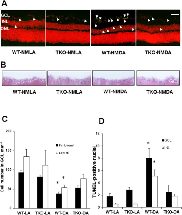
TXNIP deletion prevents long-term neuronal death and further RGC loss. (A, C) Representative images and statistical analysis of NMDA-induced neuronal death indicating significant increases in TUNEL-labelled cells (green nuclei, white arrowheads) 2 weeks post-injection in WT mice compared with TKO mice. Apoptotic cells were localized mostly to the GCL, and to a lesser extent INL and occasionally in the outer nuclear layer (ONL). Sections were counterstained with propidium iodide to label cell nuclei. Images are taken from the central region of the retina. *Significant difference as compared with the rest of the groups at P ≤ 0.05 (n = 5–7 per group, 200× magnification, scale bar is 20 μm). (B, D) Representative images and statistical analysis of the number of nuclei in the GCL from the long-term study (2 weeks post-injection) showing exacerbated cell loss (∼75%) in the GCL in WT but not in TKO mice after NMDA injection (n = 4–6 per group). *Significant difference as compared with the rest of the groups.
