FIGURE 1:
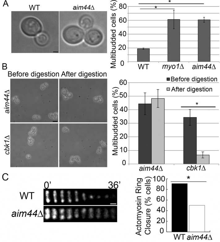
aim44∆ cells have a defect in contractile ring closure during cytokinesis. (A) Wild-type and aim44∆ cells were grown to late log phase (OD600 = 1.5) in SC glucose-based medium at 30°C, and the percentage of cells in multibudded clusters was determined. Left, transmitted-light image of wild-type cell with a single bud and an aim44∆ cell with multiple buds. Right, quantitation of the multibudded phenotype in wild-type, myo1∆, and aim44∆ cells. myo1Δ cells, which have defects in contractile ring constriction, exhibit higher levels of multibudded cells than do wild-type cells (p = 0.006). The aim44Δ cells show a statistically significant increase in the level of multibudded cells over wild type (p = 4.0 × 10⁻⁵). Error bars show SDs from three independent experiments. n ≥ 100 cells/strain per experiment. Scale bar, 1 μm. (B) The percentage of multibudded cells in aim44∆ and cbk1∆ cells was determined before and after treatment with Zymolyase 20T (0.1 mg/ml for 10 min at room temperature). Left, phase-contrast images of aim44∆ and cbk1∆ cells before and after Zymolyase treatment. Right, quantitation of multibudded phenotype before and after treatment. Zymolyase treatment results in cell separation in the cbk1∆ septation mutant (p = 0.002) but not in aim44∆ cells (p = 0.5). Errors bars show SDs from n > 800 cells/strain. Scale bar, 5 μm. (C, D) Wild-type and aim44∆ cells expressing MYO1 C-terminally tagged at its chromosomal locus with GFP were grown to mid log phase and synchronized in G1 phase by incubation with pheromone (10 μM α-factor) for 2 h at 30°C. Cells were then washed and placed in fresh media. The contractile ring was visualized beginning 60 min after release from G1 arrest by time-lapse imaging at 4-min intervals over a 40-min period. (C) Montage of the contractile ring in single cells over time. In wild-type cells (top), contractile ring closure is complete within 10 min. In contrast, in the aim44∆ cell shown, the contractile ring does not close during the 40-min imaging period (bottom). Scale bar, 0.3 μm. (D) Quantitation of the number of wild-type and aim44∆ cells that exhibit contractile ring closure (n = 52 and 66 for wild-type and aim44∆ cells, respectively; p = 7 × 10⁻⁷, chi-squared test). Results are pooled from three independent time-lapse imaging experiments.

