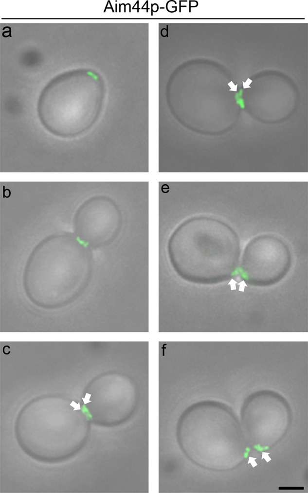
FIGURE 2:

Aim44p-GFP localization throughout the cell cycle. Cells expressing AIM44 tagged at its chromosomal locus with GFP (green) were grown as for Figure 1 and imaged by fluorescence and phase contrast microscopy. Micrographs of the fluorescence images superimposed on transmitted-light images depict representative cells at different stages in the cell division cycle. Aim44p-GFP is recruited to the selected bud site, where it forms a ring structure (a). As the bud emerges and grows, Aim44p-GFP localizes to a single ring (b) and later to a double ring (c and d, highlighted with white arrows) at the bud neck. Finally, Aim44p persists as a ring on newly separated mother and daughter cells (e and f, highlighted with white arrows). Scale bar, 1 μm (bottom right).
