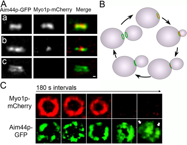
FIGURE 4:
Localization of Aim44p-GFP and Myo1p-mCherry during cell division in wild-type cells. AIM44 and MYO1 were tagged with GFP and mCherry, respectively, at their chromosomal loci. Representative images show the bud neck of mid-log-phase cells imaged by fluorescence microscopy at different points throughout the cell division cycle. (A) Projections of deconvolved z-series showing side views of Aim44p-GFP (green) and Myo1p-mCherry (red). Single rings formed by Aim44p-GFP and Myo1p-mCherry characteristic of early bud growth (a). Aim44p-GFP remains a single ring that colocalizes with the actomyosin ring through most of the cell division cycle. During contractile ring closure, the diameter of the actomyosin ring decreases (b). However, the Aim44p ring does not contract. Aim44p forms a double ring after contractile ring closure (c). Scale bar, 0.3 μm. (B) Schematic of the localizations of Aim44p-GFP (green) and the actomyosin contractile ring (red) throughout the cell division cycle. (C) Cells expressing Myo1p-mCherry and Aim44p-GFP were treated with pheromone as for Figure 1, immobilized in a microfluidic chamber such that the mitotic rings were localized parallel to the microscope stage, and imaged 60–90 min after release from pheromone-induced G1 arrest. The z-series were obtained every 3 min to observe ring dynamics before, during, and after contractile ring closure. The Myo1p-mCherry ring contracts, but the Aim44p-GFP ring does not. After completion of contractile ring closure, Aim44p-GFP forms two rings (marked by two white arrows).

