FIGURE 2:
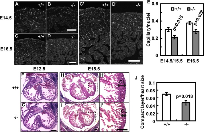
Cardiovascular defects in Borg5-null embryos. (A–D) Borg5 deletion resulted in the reduction of PECAM-1–positive microvessels in the E14.5 and E16.5 hearts. (C′) and (D′) Enlarged regions from (C) and (D), respectively. Scale bars: 100 μm. (E) Quantification of capillary density. Five mutant animals and four wild-type littermate controls at E14.5/E15.5 or E16.5 were analyzed. (F–I) Histology of wild-type and Borg5-null hearts from E12.5 and E15.5 littermates, showing an overall thinner compact layer (CL) in the E15.5 mutant heart compared with the wild-type. (H′) and (I′) Enlarged regions from (H) and (I), respectively, showing the compact layer (CL) of the left ventricles. Scale bars: 200 μm. (J) Quantification of the thickness of ventricular walls (compact layer) normalized by the heart size measured by the longest axis in E15.5 embryos. Four Borg5-null embryos and four wild-type littermate controls were analyzed. Error bars: SEM. Statistical analysis was done by two-tailed Student's t test.

