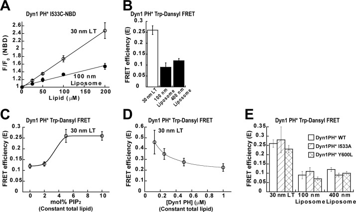
FIGURE 2:
Dyn1PH is an assembly-independent sensor of membrane curvature. (A) VL1 membrane insertion of the isolated Dyn1PH monomer (0.5 μM Dyn1PH* I533C-NBD) into increasing concentrations of LT and liposomes containing 10 mol% PIP2. The average membrane-dependent NBD emission intensity increase is indicated as F/F0, where F0 is the emission intensity at time 0 before lipid addition and F is the intensity after 30 min of incubation. (B) Binding of 0.5 μM Dyn1PH* to lipid templates of varying membrane curvature (150 μM total lipid) detected by Trp-Dansyl FRET after 30 min of incubation. Lipid templates were labeled with 10 mol% Dansyl-PE. FRET efficiency (E) was calculated according to Materials and Methods. (C) Binding of 0.5 μM Dyn1PH* to LT (150 μM constant total lipid) containing increasing mol% of PIP2 detected by Trp-Dansyl FRET. (D) FRET-detected binding of increasing concentrations of Dyn1PH* to LT (150 μM constant total lipid). (E) Binding of 0.5 μM Dyn1PH I533A (VL1) or Dyn1PH Y600L (VL3) to lipid templates of varying membrane curvature (150 μM total lipid) measured by Trp-Dansyl FRET relative to Dyn1PH WT as shown in B. All steady-state data here and in subsequent figures are averages ± SD (n ≥ 3).

