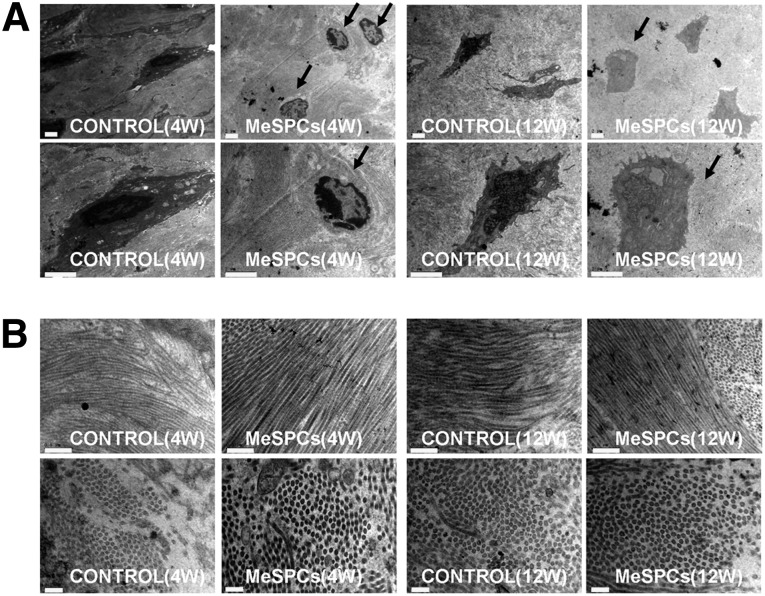
Figure 4.
The result of meniscus repair after intra-articular injection of human MeSPCs (hMeSPCs). Shown is transmission electron microscopy imaging of typical cells (A) (black arrows) and collagen fibrils (B) in the control group and hMeSPC-treated group at 4 and 12 weeks postimplantation. Scale bars = 5 µm (A), 0.5 µm ([B], top panels), and 0.2 µm ([B], bottom panels). Abbreviations: MeSPCs, meniscus-derived stem/progenitor cells; W, weeks.

