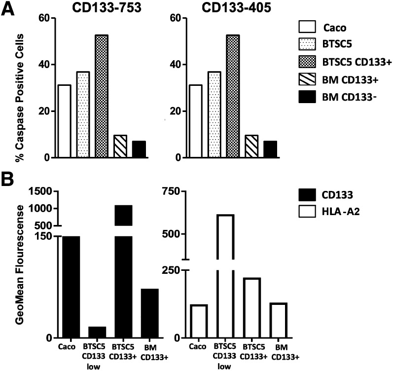
Figure 4.
Killing and characterization of CD133 expressing cells. (A): Specific killing of target CD133+ cells. CD133-753 and CD133-405 CTLs from donor H16C were cocultured with the brain tumor-derived stem cell line BTSC5 or CD133+ enriched BTSC5 cells as targets at a 20:1 ratio. The CD133+ colon cancer cell line Caco2 and BM CD133+ and CD133− cells were used as controls. After overnight incubation, caspase 3 staining was analyzed by intracellular staining. Results show the percentage of each target cell population expressing caspase 3. (B): Comparative expression of HLA and CD133 protein. HLA and CD133 expression was assessed by flow cytometry on the indicated cell populations. BM CD133+ cells were enriched from human bone marrow. BTSC5 cells were sorted by fluorescence-activated cell sorting based on CD133 expression (high or low) and cultured for 2 weeks previous to staining. Abbreviations: BM, bone marrow; HLA, human leukocyte antigen.

