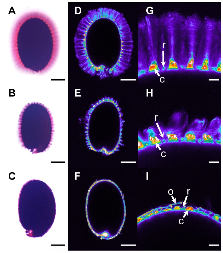
Figure 4. Adherent mucilage width and cellulose labelling are reduced in floating mucilage-releasing (FMR) accessions.

Ruthenium red staining of pectins (A) to (C) and Pontamine S4B staining of cellulose (D) to (I) in the adherent mucilage released from imbibed seeds of wild-type Col-0 (A), (D) and (G), cesa5-1 (B), (E) and (H), and the FMR accession Rak-1 (C), (F) and (I). (G) to (I) are magnifications of regions in (D) to (F), respectively. Images (D) to (G) are shown using the Rainbow2 look-up table. Bars = 150 µm (A) to (F) or 20 µm (G) to (I). C, Columella; o, outer cell wall; r, radial cell wall.
