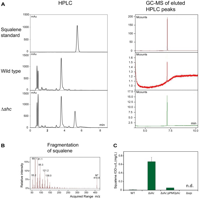
Figure 3. Analysis of squalene accumulation.
Squalene was extracted from wild type and Δshc cultures and the extracts analyzed by HPLC using a squalene standard for identification and quantification of the squalene peak. The identity of the squalene peak was confirmed using GC-MS. (A) HPLC chromatograms (left panels) and GC-MS chromatograms (right panels) for the detection of squalene. A squalene standard (top) was compared with cell extracts from wt (middle) and Δshc mutant (bottom). (B) Fragmentation of squalene standard detected using GC-MS (C) Comparison of squalene content between wild type, Δshc, Δshc:pPMQshc complemented strain, and Δsqs cells. n.d. = no squalene detected.

