
Figure 3.
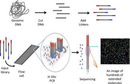
Schematic of 1 form of NGS. The process starts by randomly cutting genomic DNA (or cDNA) into short fragments (a few hundred base pairs in length). Oligonucleotide linkers are added to the fragments to generate a library in vitro. Libraries are introduced into a microscope slide with flow channels containing complementary oligonucleotides on the surfaces of the channel to ones on the libraries, thus allowing hybridization to attach millions of individual molecules to discrete locations on the slide. In situ PCR is performed to copy the individual fragments of the library to enhance sequencing detection. Single-base extension by a DNA polymerase with all 4 dye terminators extends the sequence 1 base. The image of the base extension is captured. This cycle and is repeated a 100 times from 1 end of the molecule and 100 times from the other.