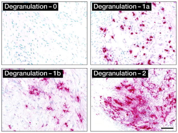
Figure 2.
Immunohistochemical analysis of eosinophil degranulation (Edgn): representative photomicrographs (400×hpf [0.29 mm2 field of view]) for each level of degranulation as determined by EPX-mAb–based immunohistochemistry (magenta staining cells and extracellular matrix regions). Scale bar = 50 μm.

