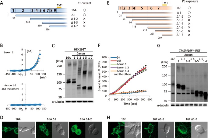
FIGURE 3.
N-terminal deletion of TMEM16A and -16F. A–D, effect of N-terminal deletion on the Cl− channel activity of TMEM16A. A, N-terminal deletion mutants of TMEM16A are shown schematically. Numbers in the top row and at the bottom indicate the exon number and the amino acid position where the deletion mutants start, respectively. TM1, first transmembrane region. The mutants that showed Cl− channel activity are marked by ○, and the mutants that did not are marked by ×. B, membrane currents at the indicated voltage pulses (mV) were measured by whole-cell patch clamp analysis of 293T cells transfected with the expression vector for the deletion mutant (Δexon 1, Δexon 1–2, Δexon 1–5, or Δexon 1–7 of TMEM16A). C, 293T cells that had been transfected with the expression vector for TMEM16A or its N-terminal deletion mutants were separated by 5–10% SDS-PAGE and analyzed by Western blotting with an anti-FLAG mAb. D, HEK293T cells were transfected with the expression vectors for GFP-tagged wild-type or deletion mutants (Δ1 and Δ1–2) of TMEM16A and observed using a confocal microscope. E–H, effect of N-terminal deletion on the phospholipid scramblase activity of TMEM16F. E, N-terminal deletion mutants of TMEM16F are shown. Numbers in the top row and at the bottom indicate the exon number and the amino acid position where the deletion mutants start, respectively. The mutants that showed the scramblase activity are marked by ○, and the mutants that did not are marked by ×. F, Cy5-Annexin V binding to the A23187-treated TMEM16F−/− IFET transformants expressing the full-length TMEM16F or its deletion mutant (Δexon 1, Δexon 1–2, Δexon 1–3, Δexon 1–4, Δexon 1–5, Δexon 1–6, or Δexon 1–7) was followed by FACSCalibur for 9 min and expressed as mean fluorescence intensity (MFI). G, Western blotting of TMEM16F−/− IFET transformants expressing TMEM16F or its deletion mutants. Cell lysates were separated by 5–10% SDS-PAGE and analyzed with an anti-FLAG mAb. H, HEK293T cells were transfected with the expression vectors for GFP-tagged wild-type or deletion mutants (Δ1–2 and Δ1–3) of TMEM16F and observed using a confocal microscope. Error bars represent S.D.

