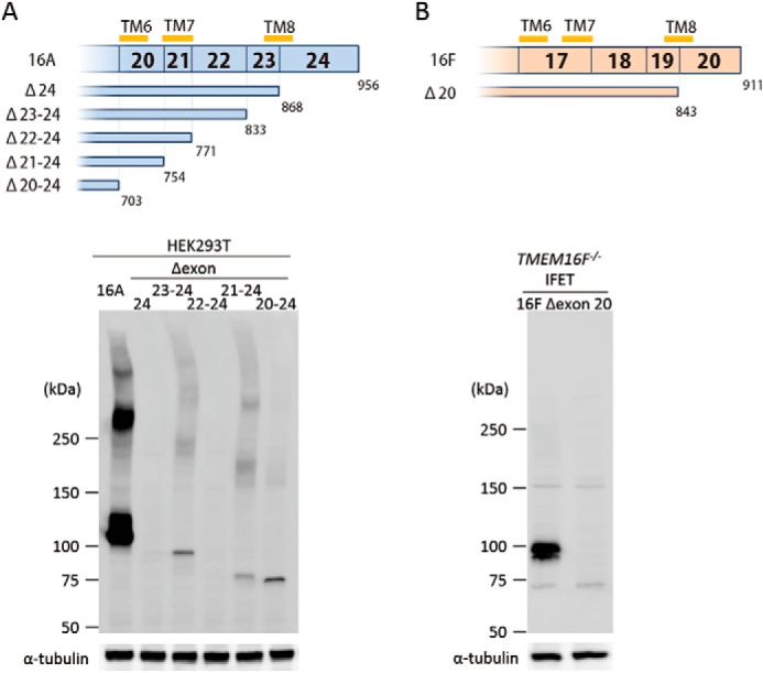
FIGURE 4.

C-terminal deletion of TMEM16A and -16F. A, effect of C-terminal deletion on the expression of TMEM16A. In the upper panel, structures of the C-terminal deletion mutants of TMEM16A are schematically shown. Numbers in the top row indicate the exon number. TM6, TM7, and TM8 represent the sixth, seventh, and eighth transmembrane regions. Numbers at the bottom indicate the amino acid position where the deletion mutants end. In the lower panel, the cell lysates from 293T cells that had been transfected with the vector for full-length 16A or its C-terminal deletion mutants (Δexon 24, Δexon 23–24, Δexon 22–24, Δexon 21–24, and Δexon 20–24) were separated by 5–10% SDS-PAGE and analyzed by Western blotting with an anti-FLAG mAb. As a control, Western blotting was performed with an anti-α-tubulin mAb, and the results are shown at the bottom. B, effect of C-terminal deletion on the expression of TMEM16F. The structure of the C-terminal deletion mutant of TMEM16F is shown. Numbers in the top row indicate the exon number. TM6, TM7, and TM8 represent the sixth, seventh, and eighth transmembrane regions. Numbers at the bottom indicate the amino acid position where the deletion mutant ends. In the lower panel, the lysates from TMEM16F−/− IFET transformants expressing the full-length 16F or its C-terminal deletion mutant (Δexon 20) were separated by 5–10% SDS-PAGE and analyzed by Western blotting with an anti-FLAG mAb. As a control, Western blotting was performed with an anti-α-tubulin mAb, and the results are shown at the bottom.
