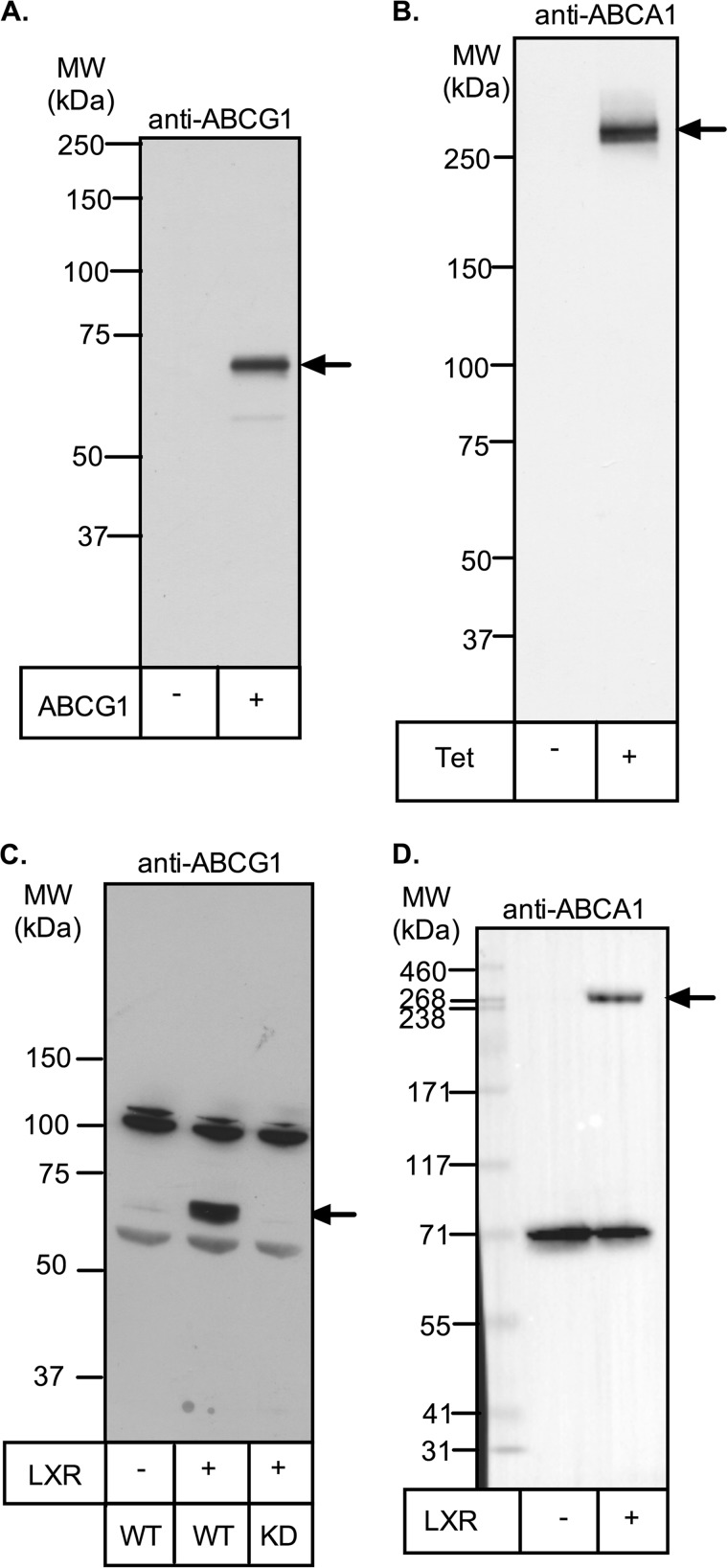
FIGURE 1.
Detection of ABCA1 and ABCG1 by Western blot. A, cell lysate (10 μg of protein) from CHO cells stably expressing ABCG1-678 was separated by SDS-PAGE (7% Tris acetate gel) and subjected to Western blotting for ABCG1 (Novus rabbit polyclonal antibody; 1:2500) as described under “Experimental Procedures.” B, ABCA1-CHO cells were preincubated overnight with or without tetracycline (1 μg/ml) to induce ABCA1 expression. Cell lysates (30 μg of protein) were separated by SDS-PAGE and visualized by Western blotting for ABCA1 (Life Research mouse monoclonal; 1:2500) as described under “Experimental Procedures.” C and D, THP-1 cells were preincubated for 24 h with (second lane) or without (first lane) T0901317 (1 μm) to induce ABCA1 and ABCG1 expression. Cell lysates (15 μg of protein) were then separated by SDS-PAGE and visualized by Western blotting for either ABCG1 (Novus rabbit polyclonal antibody; 1:2000; C) or ABCA1 (Novus rabbit polyclonal antibody; 1:2000; D) as described under “Experimental Procedures.” Positions of molecular mass markers are shown, and arrows indicate the bands of ABCA1 or ABCG1. KD, THP-1 cell clone in which ABCG1 is stably silenced (19). MW, molecular mass.

