FIGURE 4.
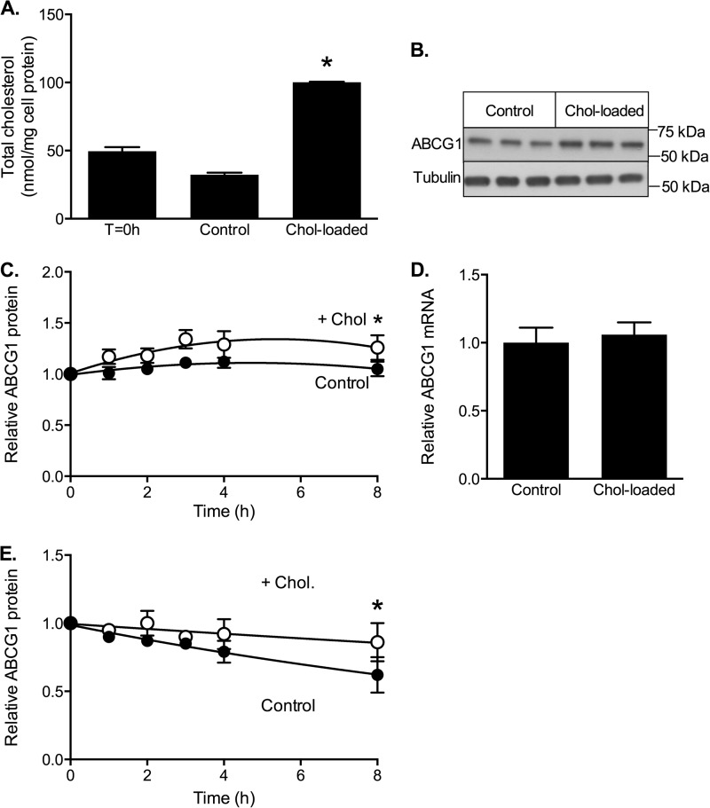
Effects of cholesterol on stability and degradation of ABCG1-666. CHO cells stably expressing ABCG1-666 were incubated for up to 8 h in either low cholesterol (control) medium (0.5% (v/v) FBS) or cholesterol-enriched medium (0.5% FBS supplemented with cholesterol/CD (20 μg cholesterol/ml)) (Chol-loaded). A, cholesterol content of cells before (T = 0 h) and after incubation for 8 h with or without cholesterol. The data are means ± S.D. of triplicate measurements in a single representative experiment. B, Western blot showing cellular ABCG1 protein after 8 h with or without cholesterol. C, time course of ABCG1 protein expression during incubation with (white circles) or without (black circles) cholesterol. The data are means ± S.D. of five independent experiments. D, ABCG1 mRNA levels measured after 8 h of incubation with or without cholesterol. The data are means ± half-range of two separate experiments, each performed in triplicate. E, decline in ABCG1 protein after addition of cycloheximide (10 μg/ml) in the presence (white circles) or absence (black circles) of cholesterol. The data are means ± S.D. of five independent experiments. *, significant difference (p < 0.05) relative to control cells.

