
FIGURE 3.
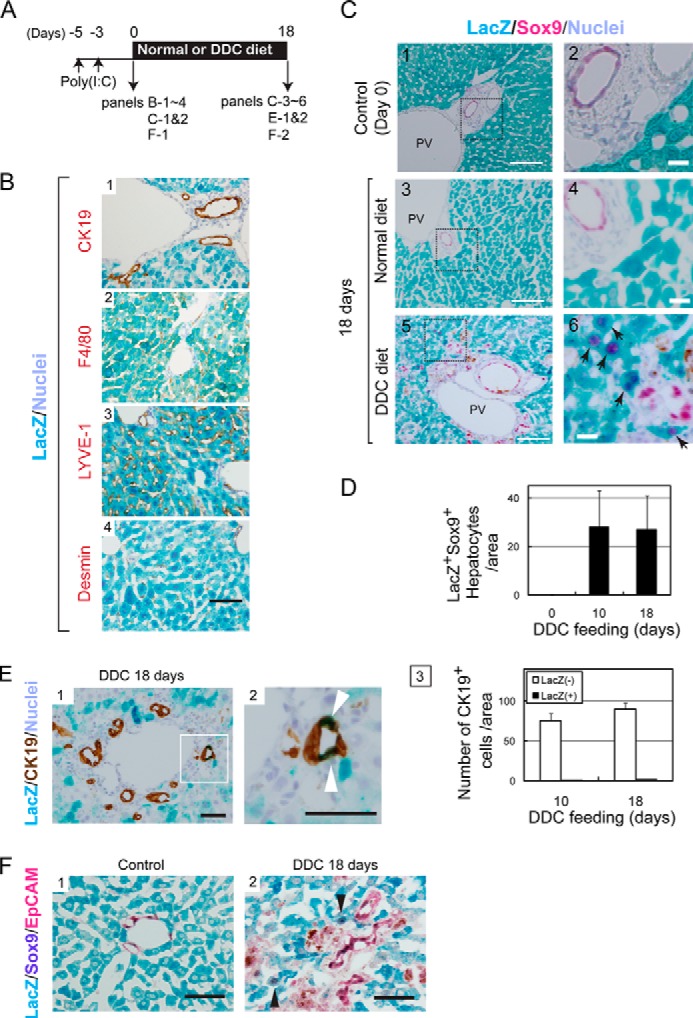
Mature hepatocytes are the origin of Sox9+ biphenotypic cells. A, the time course of the labeling of MHs in Mx1-Cre:ROSA mice and the induction of liver injury. B, specificity of hepatocytes labeling in Mx1-Cre:ROSA mice. Hepatocytes were labeled with LacZ after poly(I:C) injection. On the other hand, CK19+ cholangiocytes, LYVE-1+ sinusoidal endothelial cells, F4/80+ Kupffer cells, and Desmin+ stellate cells were not labeled with LacZ in Mx1-Cre:ROSA mice injected with poly(I:C). Bar represents 50 μm. C, LacZ+ hepatocytes convert to Sox9+ cells. Before DDC injury, LacZ staining is limited to MHs (panels 1 and 2). After feeding mice with the DDC diet for 10 and 18 days, LacZ+Sox9+ cells appear near the portal vein (PV) (arrows in panels 4 and 6). Bars in panels 1, 3, and 5 represent 100 μm, while those in panels 2, 4, and 6 represent 20 μm. D, the number of LacZ+Sox9+ cells with hepatocyte morphology. The number of LacZ+Sox9+ cells showing hepatocyte morphology was counted on liver sections of normal and DDC diet-fed mice. Error bars represent S.E. E, CK19+LacZ+ cells emerge in DDC-injured livers of Mx1-Cre:ROSA mice. After 18 days of DDC injury, some of LacZ+ cells are incorporated to ductular structures as CK19+ cells (white arrowheads). The number of CK19+LacZ+ cell is slightly increased between 10 and 18 days of DDC injury. At day 18, it represents about 2% of CK19+ cells. Bars in panels 1 and 2 are 50 μm. F, LacZ+Sox9+ cells emerging in DDC-injured liver of Mx1-Cre mice are negative for EpCAM. In the control liver, LacZ+ cells express neither Sox9 nor EpCAM (panel 1). After DDC injury, LacZ+Sox9+ biphenotypic cells, which are negative for EpCAM, are observed (arrowheads in panel 2). EpCAM and Sox9 were visualized with Permanent Red and 5-bromo-4-chloro-3-indolyl phosphate, respectively. Bars represent 50 μm.