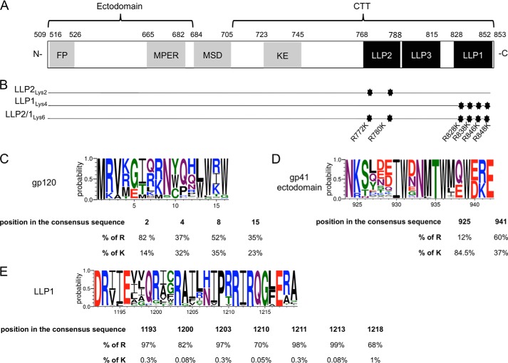
FIGURE 1.
Representation of the CTT mutations and amino acids conservation. A, schematic representation of gp41 from the 89.6 virus strain of HIV-1. The gp41 subunit is divided into three main parts: an ectodomain, a MSD, and a CTT. The residue numbering is based on the gp160 protein. gp120 subunit is not represented. FP, fusion peptide; MPER, membrane proximal external region; KE, Kennedy epitope. B, the most conserved LLP arginines were mutated into lysines. Three mutants were constructed containing two mutations in LLP2 (LLP2Lys2), four mutations in LLP1 (LLP1Lys4), and the third mutant contains all six mutations (LLP2/1Lys6). C–E, logo diagrams representing the relative frequency (y axis) for each amino acid of selected representative regions of gp120 (C), the ectodomain of gp41 (D), and LLP1 (E). Below each logo diagram are represented the percentages of arginine or lysine residue frequencies in the alignment at the indicated position.

