
FIGURE 1.
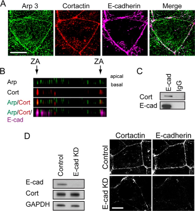
Cortactin accumulates with Arp2/3 at apical E-cadherin junctions. A, maximum projection view from a confocal stack of Arp3, cortactin, and E-cadherin immunostaining in Caco-2 cells. Scale bar, 10 μm. B, Z-section of image in A showing that Arp3 (Arp, green) and cortactin (cort, red) co-localize at the apical regions (ZA) of cell-cell contacts, as marked by E-cadherin (E-cad, magenta). C, E-cadherin or control IgG immunoprecipitates from Caco-2 cells immunoblotted for cortactin (Cort) or E-cadherin (E-cad). D, impact of E-cadherin on cortactin localization. Cells treated with E-cadherin siRNA to KD E-cadherin were analyzed by Western blotting (left panel) and immunofluorescence (right panel) for cortactin and E-cadherin; GAPDH was used as a loading control. Junctional cortactin was lost, despite stable cellular expression, in E-cadherin KD cells. Scale bar, 10 μm.