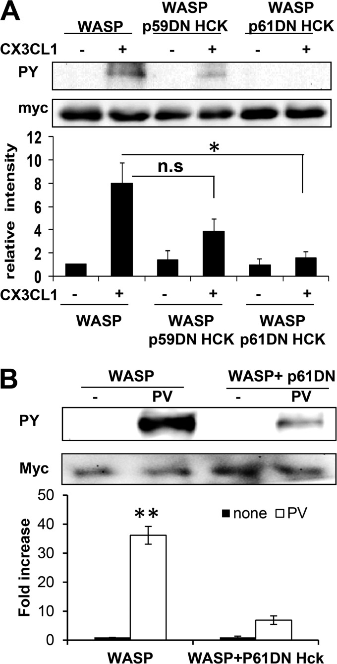
FIGURE 6.
The p61Hck isoform regulates WASP tyrosine phosphorylation. A, cells were transfected with either Myc-WASP alone or with dominant-negative form of p61Hck (p61DN) or p59Hck (p59DN) and then stimulated with CX3CL1 for 1 min, and then WASP was immunoprecipitated using a Myc antibody, followed by Western blotting with the HRP-conjugated phospho-tyrosine (PY) and Myc antibody as a control. Blots were quantified by densitometry and normalized to amount of WASP. A representative blot is shown, and data are expressed as the fold increase compared with WT prior stimulation. Shown are means ± S.E. (n = 3). B, cells were transfected with either Myc-WASP alone or with dominant-negative p61Hck (p61DN) and stimulated with pervanadate (PV) to induce maximal tyrosine phosphorylation. The graph shows the fold change in WASP phosphotyrosine (n = 3), mean ± S.E. **, p < 0.01 compared with untreated cells (none). n.s, not significant.

