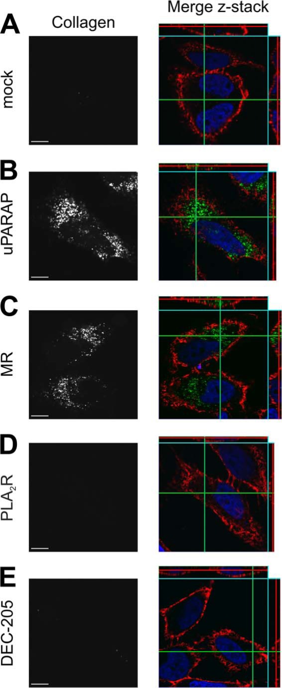
FIGURE 4.

Endocytosis of fluorescent gelatin in uPARAP and MR-transfected cells. Internalization of fluorescently labeled gelatin by HeLa cells transfected with empty vector (mock, A), uPARAP (B), MR (C), PLA2R (D), and DEC-205 (E). Cells were incubated for 16 h with fluorescent collagen ligand (20 μg/ml, right panels, green). Cell nuclei were stained with Hoechst (right panels, blue) and cell membranes with wheat germ agglutinin (right panels, red). Z-stacks were collected using a confocal microscope. Left panels show the signal from fluorescent collagen in gray scale in a single plane. Size bar:10 μm.
