FIGURE 8.
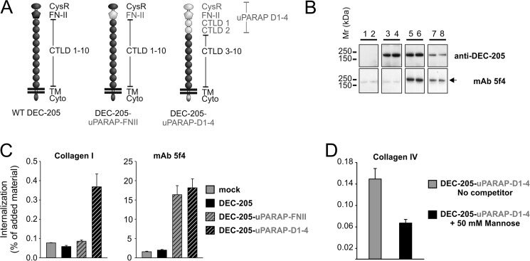
Gain of collagen internalization function in DEC-205 chimera with uPARAP D1–4 domain cassette. A, schematic overview of domain-swaps in DEC-205 chimeras (DEC-205-uPARAP-FN-II, DEC-205-uPARAP-D1-4). Inserted domains from uPARAP (gray) are highlighted to distinguish them from remaining DEC-205 domains (black). B, Western blot analysis of HEK-293T cells transfected with receptor chimeras using rabbit mAb against C-terminal DEC-205 or mAb 5f4 as primary antibody. Lanes 1 and 2: mock, lanes 3 and 4: DEC-205, lanes 5 and 6: DEC-205-uPARAP-FN-II, lanes 7 and 8: DEC-205-uPARAP-D1–4. Note that mAb 5f4 detects the FN-II domain from uPARAP in the two DEC-205 chimeras of ∼205 kDa (marked by a black arrowhead). Experimental conditions were as described in Fig. 2. C, internalization of radiolabeled collagen type I (100 ng/ml, left panel) and control ligand (100 ng/ml mAb 5f4, right panel) by HEK-293T cells transfected with DEC-205 wt and chimeras in the presence of E64d (20 μm). D, internalization of radiolabeled collagen type IV (100 ng/ml) by HEK-293T cells transfected with DEC-205-uPARAP-D1-4 in the absence or presence of 50 mm mannose. Experimental conditions and data presentation were as described for Fig. 6 (C and D).

