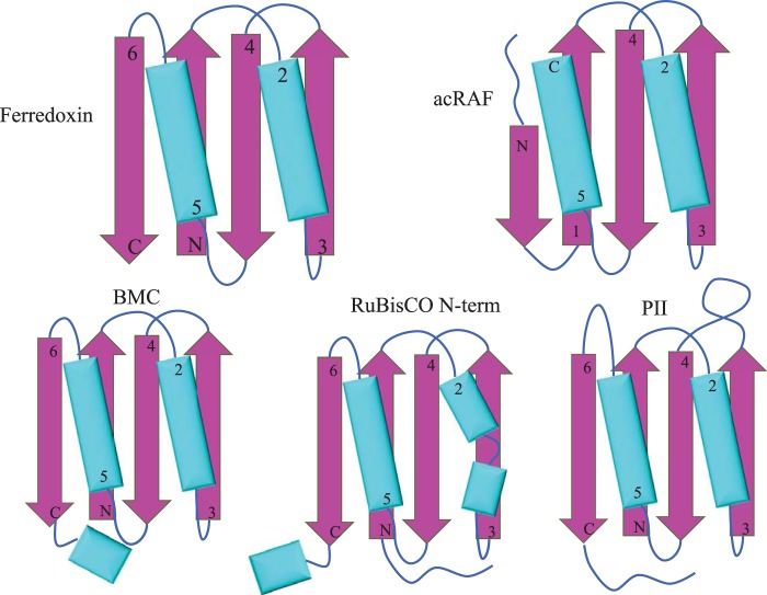
FIGURE 4.
Ferredoxin-like protein folds associated with carboxysomes. Simplified models of secondary and tertiary structural motifs of select carboxysome-associated proteins. α-Helices are colored cyan and β sheets are colored magenta. Top left, ferredoxin. Top right, PCD/acRAF. Bottom left, BMC (bacterial microcompartments shell proteins). Bottom center, N-terminal domain of the RuBisCO large subunit. Bottom right, nitrogen regulatory PII family protein (encoded near several chemoautotrophic α-carboxysome operons).

