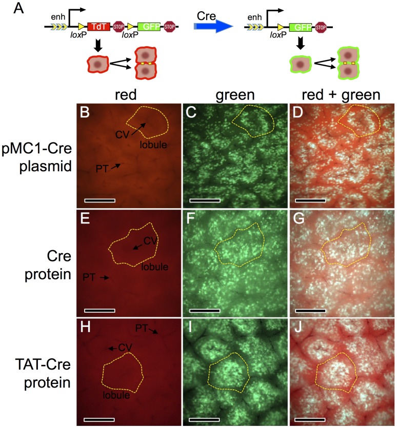
Figure 1. Cre activity in mouse hepatocytes following hydrodynamic delivery of Cre-expression plasmid or recombinant Cre proteins.
Panel A, schematic of the ROSAmT−mG double-fluorescent reporter allele [2] used in this study. In the Cre-naïve state (left), the outer membranes of all cells fluorescent red. Following Cre exposure (right), the outer membrane of cells and all of their descendents fluoresce green. Panels B–J, young adult ROSAmT−mG/mT−mG mice were given hydrodynamic injections of lactated Ringer’s saline containing 25 µg of pMC1-Cre plasmid (panels B–D), 2 nmol of recombinant His-tagged Cre protein (panels E–G), or 2 nmol of recombinant His-tagged TAT-Cre protein (panels H–J). Mice were sacrificed ≥7 d later and whole livers were photographed using an epifluorescent dissecting steromicroscope. Panels B–D are the same frame photographed under different fluorescent channels, as are E–G and H–J. A representative lobule and representative central veins (CV) and portal triads (PT) are indicated. Scale bars = 0.5 millimeter.

