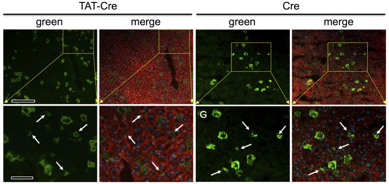
Figure 3. Hydrodynamic delivery of Cre or TAT-Cre protein targets hepatocytes.
Cryosections of livers harvested from mice as in Fig. 1 were photographed as in Fig. 2. The yellow dotted boxes in upper panels correspond to the enlarged images in lower panels, as indicated. Regions containing regions of green membrane that appeared smaller than hepatocytes are shown (white arrows) The merged images revealed that these small regions of green membrane generally did not contain within them a DAPI-stained (blue) nucleus suggesting these were not small non-hepatocyte cells, but rather, are the “tips” or “edges” of large hepatocytes whose bulk lay in adjacent sections, as further evaluated in Fig. 4. Scale bars: in upper panels = 50 µmeter; in lower panels = 16 µmeter.

