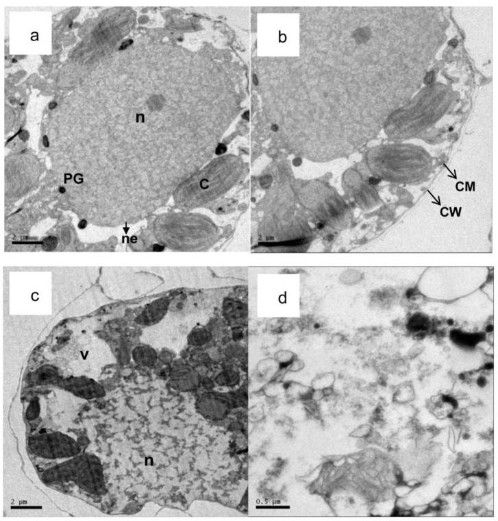
Figure 8. Transmission electron micrographs of the lysing process in A. tamarense treated with palmitoleic acid (40 μg/mL).
(a,b) control cells of A. tamarense (×1000); (c) a damaged A. tamarense cell after 2 d, (×1000); (d) a damaged A. tamarense cell after 5 d, (×1000). (CM: cell membrane; CW: cell wall; V: vacuole; n: nucleus; C: chloroplast; ne: nuclear envelope and PG: plastoglobule.).

