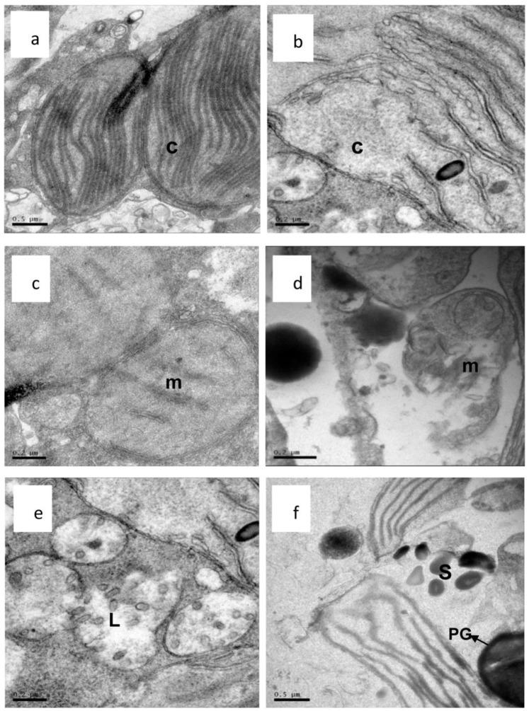
Figure 9. Ultrastructure of A. tamarense cells stressed by palmitoleic acid (40 μg/mL).
(a,b) an intact chloroplast and a damaged chloroplast after 3 d (×50,00); (c,d) an intact mitochondrium and a damaged mitochondrium after 3 d (×15,000); (e,f) a damaged lysosome after 3 d and randomly distribution of the starch grains and plastoglobule (×15,000). (C: chloroplast; m: mitochondria; L: lysosomes; PG” plastoglobule and S: starch grains.).

