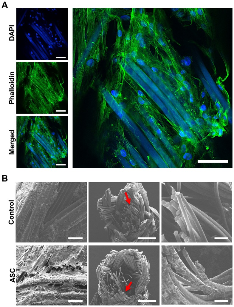
Figure 2. Cellular distribution and attachment in the suture.
ASCs were filled into biodegradable sutures. Laser scanning microscopy (LSM, A) shows attached cells (DAPI, blue) along the suture surface interacting with each other (phalloidin, green) and the suture itself (DAPI, blue/phalloidin, green). The suture material is autofluorescent. As observed by scanning electron microscopy (SEM, B), cells were distributed throughout the surface (left) and the inner filaments (middle, right) of the suture. Scale bars represent 50 (A and B left), 500 (B, middle), and 100 μm (B, right).

