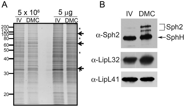
Figure 1. Virulent leptospires become mammalian host-adapted during growth within dialysis membrane chambers.
Representative whole cell lysates of leptospires cultivated to late-logarithmic phase in EMJH medium at 30°C in vitro (IV) and within dialysis membrane chambers (DMC) implanted into the peritoneal cavities of female Sprague-Dawley rats. (A) Lysates were loaded according to the numbers of leptospires (5×10⁶ per lane) or total protein (5 µg per lane) and stained with SYPRO Ruby gel stain. Arrows and asterisks are used to highlight examples of polypeptides whose expression appears to be increased or decreased, respectively, within DMCs compared to in vitro. Molecular mass markers are indicated on the left. (B) Immunoblot analyses using rabbit polyclonal antisera directed against Sph2 [34], LipL32 [38] and LipL41 [39]. An arrow is used to indicate a band of the predicted molecular mass for SphH, a second, closely-related sphingomyelinase in L. interrogans recognized by antiserum directed against Sph2 [34], [37].

