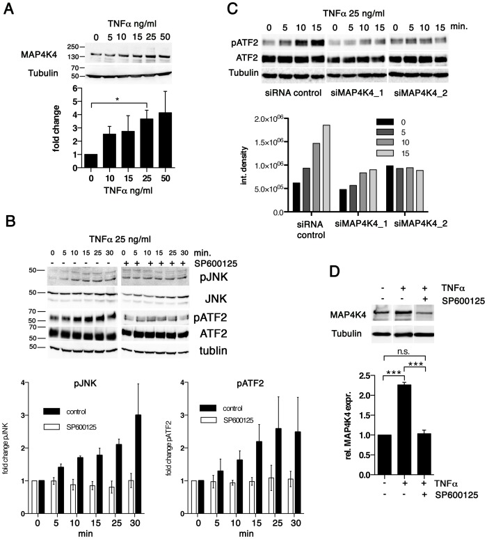
Figure 4. TNFα promotes increased MAP4K4 protein expression through the JNK pathway.
A) IB analysis of cells treated for 24 h with increasing concentrations of TNFα using antibodies against MAP4K4 or tubulin; Below: quantification of integrated densities of bands (means −/+ SDs, normalized to tubulin). B) Time course IB analysis of lysates of cells treated with 25 ng/ml TNFα using antibodies against proteins indicated. Right: IB analysis of effect of JNK inhibitor SP600125 (10 ng/ml, added 2 h before TNFα treatment) on TNFα-induced JNK pathway activation. Bar diagram: Means and SDs of pJNK and pATF2 after TNFα stimulation (normalized to total JNK and total ATF2 proteins, respectively). C) Time course IB analysis of lysates of cells treated with 25 ng/ml TNFα using antibodies against proteins indicated. Cells were stimulated 24 h after transfection with siControl or siMAP4K4_1 or siMAP4K4_2. Bar diagram below shows quantification of pATF2 bands (normalized to total ATF2 protein). D) IB analysis of lysates from cells treated for 24 h−/+25 ng/ml TNFα and −/+10 ng/ml SP600125, using anti-MAP4K4 or anti-tubulin antibodies. Bar diagram below shows quantification of MAP4K4 bands (means −/+ SDs, normalized to tubulin).

