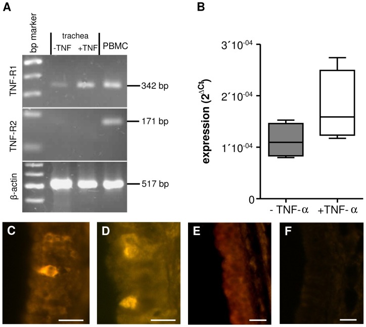
Figure 4. Expression of TNF-R1 in tracheal epithelium.
A: In RT-PCR experiments using whole tracheal preparations we found evidence for mRNA of TNF-R1 but not TNF-R2 (n = 3, bp: 100 kD). B: In tracheae previously exposed to TNF-α over 60 min we observed a non-significant increase in TNF-R1 mRNA when using qPCR (p = 0.2, n = 4). C: In addition immunoreaction against TNF-R1 was used to identify the anatomical location of the TNF-R1. Immunoreaction against TNF-R1 stained only cells within the epithelial layer. These were either found as isolated cells or as clusters of 2–4 cells (D). E: Pre-absorption of the primary antibody with TNF-R1 peptide did not stain any cells in the tracheal epithelium. F: In addition experiments omitting primary antibody were used as negative controls evoked no immunoreactive staining of epithelial structures. (Scale bars: 20 μm).

