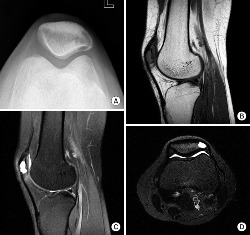
Fig. 1.
A radiograph of the left knee showed a well-defined lytic lesion adjacent to the articular surface with a sclerotic margin in the lateral aspect of the patella (A). A focal lesion with low signal intensity was observed in T1-weighted magnetic resonance imaging (MRI) (B). High signal intensity and fluid-fluid levels were also seen in T2-weighted MRI (C, D).

